♫ Dem bones, dem bones, dem dry bones.
…Now shake dem skeleton bones!The foot bone’s connected to the leg bone.
The leg bone’s connected to the knee bone.
The knee bone’s connected to the thigh bone.
Now shake dem skeleton bones!♫
The Inside Scoop on Bones
 When you think about bones, you probably conjure up visions of dinosaur skeletons or maybe a human skeleton from high school biology. But what are bones inside your body really like?
If you think the bones inside your body are dry, hard and unchanging, you’d be wrong. Your bones are alive, your skeleton is an active organ: growing and transforming throughout your life.
Parts of Bones
To understand problems with bones, you have to know the parts of the bones and their function. So here is a short review.
The outside of the bone is covered with a thin layer called the periosteum. This layer contains collagen on the outside with blood vessels that nourish the bone and nerves involved in balancing and pain reception. The inner layer of the periosteum is made up of osteoblasts (see the types of cells below.)
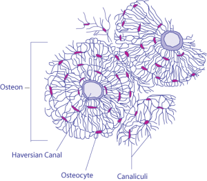 Right under the periosteum is a layer called cortical or compact bone. This is the hard smooth layer of bone. It is made up of cells, called osteocytes, surrounding canals (Haversian Canals) filled with blood vessels and nerves. One canal and the osteocytes that ring it is called an osteon. The compact layer is made up of these osteons lying parallel to each other.
Further into the bone is trabecular or cancellous bone.
Cancellous bone has many pores or holes in it, giving the appearance of a sponge. Don’t be fooled, cancellous bone it is very hard. In the pores are more blood vessels, nerves and (in flat bones like the skull) bone marrow.. 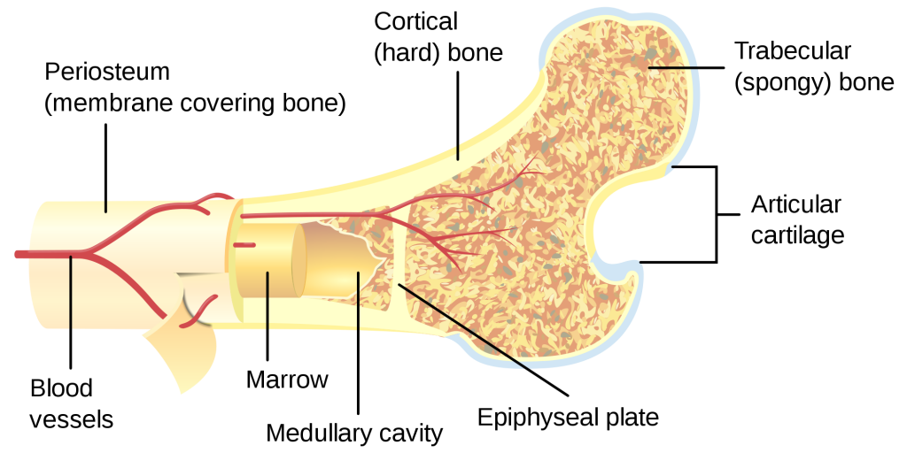 In long bones–legs, arms, jaw–like the one pictured above, the center of the bone is the bone marrow. There are 2 types of bone marrow, red and yellow. Most of the blood of the body–red blood cells, white blood cells and platelets–is made in red marrow. Yellow marrow is mostly fat cells. As an adult half of your marrow is red; the other half yellow.
Cells of the bone
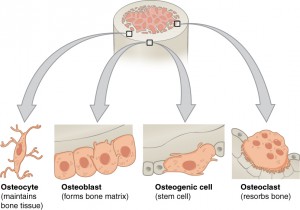
There are three types of bone cells: Osteogenic, osteoblasts, osteocytes and osteoclasts.
- Osteoblasts-These are bone forming cells. Some of these cells flatten out to line the surfaces of bones. Osteogenic cells are the precursors of osteoblasts and are also called stem cells.
- Osteocytes-Osteoblasts that have become part of the compact layer of the bone. They maintain the bone.
- Osteoclasts-These are cells that resorb bone–they break down bone.
Resorption, Bone Modeling and Remodeling
During childhood until humans are around 25 years old, our skeleton grows. Bone modeling is the term for this process. Bone is added in some places and the bone is resorbed in other places. Resorption means that the bone is broken down and the minerals go into the blood stream. The minerals are recycled to make new bone or eliminated from the body. Modeling is responsible for the skeleton changing its form.
By the time we are 25, our bodies contain the largest amount of bone mass that we will have throughout our lives. During adulthood, bone remodeling occurs. Old, mature bone tissue is replaced by new bone tissue. This cycle of maintenance helps retain bone strength.
Balance is key. Throughout the process, all the bone cells communicate with each other. Since osteoclasts resorb minerals faster than osteoblast form bone, balance can be hard to maintain.
Osteopenia and Osteoporosis
Osteopenia and subsequent osteoporosis occurs when bone remodeling is out of balance. If too much bone is resorbed, osteopenia, or bone weakening occurs. Bone loss through resorption or too little bone formation makes bones become brittle and likely to fracture. This is osteoporosis. Researchers have been exploring what causes an imbalance in osteoclastic and osteoblastic activity to occur. Hormones like estrogen, testosterone and parathyroid hormone seem to play a role. Research has found receptors on osteoblasts and osteoclasts that switch them on and off. More research is needed.
Who Is At Risk For Osteoporosis?
Women (1 in 2) are more likely than men (1 in 4) to develop osteoporosis. Certain chronic diseases increase the risk of osteoporosis. For example, both Type 1 and Type 2 Diabetes increase the risk of osteoporosis but different mechanisms are involved. Prostate cancer is a risk factor as well. Certain cancer treatments increase the risk of osteoporosis. Below is a diagram (created from information on the National Osteoporosis Foundation website) detailing some of the conditions that increase risk of osteoporosis. 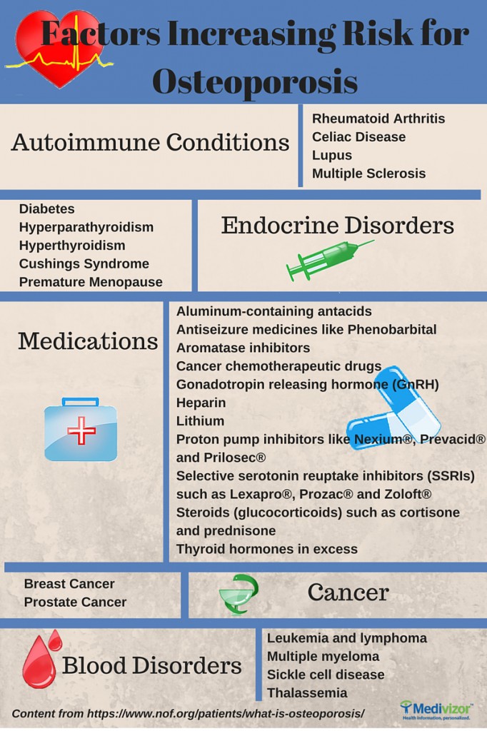
Prevention
Having a higher bone mass as a child and young adult can help to stave off bone loss in later life. To help reduce the risk of getting osteoporosis, there are several recommendations.
1) Don’t smoke.
2) Stay physically active: do weight bearing muscle strengthening exercises
4) If you drink alcohol, do so in moderation.
5) Be sure to get enough Vitamin D.
6) If you have risk factors, get a bone density test.
7) Stay alert to signs of bone loss, for example loss of height.
8) Talk to your doctor about osteoporosis.
Bones Are Alive
So, did your mom explain all of this to you? Are you ready for the bones are alive quiz? Then someone else needs to give that to you.
What’s the take-away? May is Osteoporosis Awareness Month. After age 50, everyone is at risk for osteoporosis! So drink your milk already!



Med Hypotheses. 2001 Feb;56(2):163-70.
http://www.ncbi.nlm.nih.gov/pubmed/11425281
The multifaceted and widespread pathology of magnesium deficiency.
Johnson S.
Abstract
Even though Mg is by far the least abundant serum electrolyte, it is extremely important for the metabolism of Ca, K, P, Zn, Cu, Fe, Na, Pb, Cd, HCl, acetylcholine, and nitric oxide (NO), for many enzymes, for the intracellular homeostasis and for activation of thiamine and therefore, for a very wide gamut of crucial body functions. Unfortunately, Mg absorption and elimination depend on a very large number of variables, at least one of which often goes awry, leading to a Mg deficiency that can present with many signs and symptoms. Mg absorption requires plenty of Mg in the diet, Se, parathyroid hormone (PTH) and vitamins B6 and D. Furthermore, it is hindered by excess fat. On the other hand, Mg levels are decreased by excess ethanol, salt, phosphoric acid (sodas) and coffee intake, by profuse sweating, by intense, prolonged stress, by excessive menstruation and vaginal flux, by diuretics and other drugs and by certain parasites (pinworms). The very small probability that all the variables affecting Mg levels will behave favorably, results in a high probability of a gradually intensifying Mg deficiency. It is highly regrettable that the deficiency of such an inexpensive, low-toxicity nutrient result in diseases that cause incalculable suffering and expense throughout the world. The range of pathologies associated with Mg deficiency is staggering: hypertension (cardiovascular disease, kidney and liver damage, etc.), peroxynitrite damage (migraine, multiple sclerosis, glaucoma, Alzheimer’s disease, etc.), recurrent bacterial infection due to low levels of nitric oxide in the cavities (sinuses, vagina, middle ear, lungs, throat, etc.), fungal infections due to a depressed immune system, thiamine deactivation (low gastric acid, behavioral disorders, etc.), premenstrual syndrome, Ca deficiency (osteoporosis, hypertension, mood swings, etc.), tooth cavities, hearing loss, diabetes type II, cramps, muscle weakness, impotence (lack of NO), aggression (lack of NO), fibromas, K deficiency (arrhythmia, hypertension, some forms of cancer), Fe accumulation, etc. Finally, because there are so many variables involved in the Mg metabolism, evaluating the effect of Mg in many diseases has frustrated many researchers who have simply tried supplementation with Mg, without undertaking the task of ensuring its absorption and preventing excessive elimination, rendering the study of Mg deficiency much more difficult than for most other nutrients.
Thank you for sharing this link. Kathleen
Wow, this is very comprehensive. I learned a lot about bone health. Thanks!